
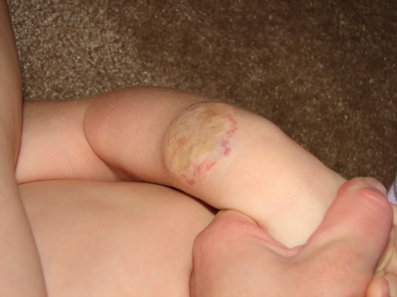
 Above is a picture of the hemangioma on The Wild Child's arm at 5 weeks old. I wasn't thinking this was going to be that big of a deal, so I actually don't have any pictures from earlier in her life. It started flat and light red, but you can see by this time, it was raised and getting dark. The part in the middle that is purple is the part that was soon to ulcerate (break open). This when I realized I needed to start taking weekly pictures to document the rapid growth.
WC's H is most likely a Superficial Hemangioma (formerly called Strawberry Hemangioma) because it is located in the surface of the skin. They are characterized by a bright red color with a well defined border. These lesions may vary greatly in size, location and shape.
PickleLovingPrincess, you mentioned a Deep Hemangioma (formerly known as Cavernous Hemangioma), which is often larger than a superficial lesion. They are located beneath the surface of the skin and are characterized by a bluish lump or mass. They are generally soft to the touch.
It is possible that WC's was a Combined Hemangioma or Mixed Hemangioma, both deep and superficial. These may appear as a lump or mass in the skin with areas of red. They are often soft. We did not, however, have any testing done to see how deep it went so I have to assume it was just a Superficial.

This picture was taken at 17 weeks of age. The ulceration was at it's worst now, but beginning to heal. This was a stressful time for us- the ulceration was very painful and although we were fortunate that her H wasn't on her face as most of them are, but it was still in an area that was easily bumped (imagine threading those little arms into a carseat.....), which would start off a 2 hours crying spell because of pain that Tylenol didn't touch.
We also had some clothing issues because anything with a wristband was a problem, so I had a difficult time figuring out what to do about winter PJs.....
It was at this point that our ped finally agreed that we needed to see a specialist. Up until now we were told "We don't do anything for those, just wait them out and they will go away on their own." (oh-so-wrong I would later find out....) To make a really long story a little shorter (you can email me for the long version if you'd like) we found a doctor in a neighboring state who was a pediatric plastic surgeon who specializes in vascular anomalies. He let us know that even if the H fully involutes, she will still be left with a cosmetic deformity because of the significant scar tissue that formed because it was an open wound for so long. So our plan at this point is to do a scar revision surgery after her third birthday.
This picture was taken when she was about 18 months old. The area in the middle that has a different texture is the scar tissue.
I do have a wealth of links and support groups/forums if anyone is ever in need of them. Just let me know and I'll be glad to share my resources!




 NARS Lip Stain Gloss


